Breast CT: Revolutionary Technology for Breast Imaging
April 21, 2022
April 21, 2022
Customized data transfer solutions for the medical industry

Today's Challenge
In today's medical market, the traditional mammogram can cause discomfort and expose patients to a low dose of radiation. Patients who wear pacemakers or have older silicone implants can also be denied access due to the nature of the breast examination.
Moog GAT Has A Solution
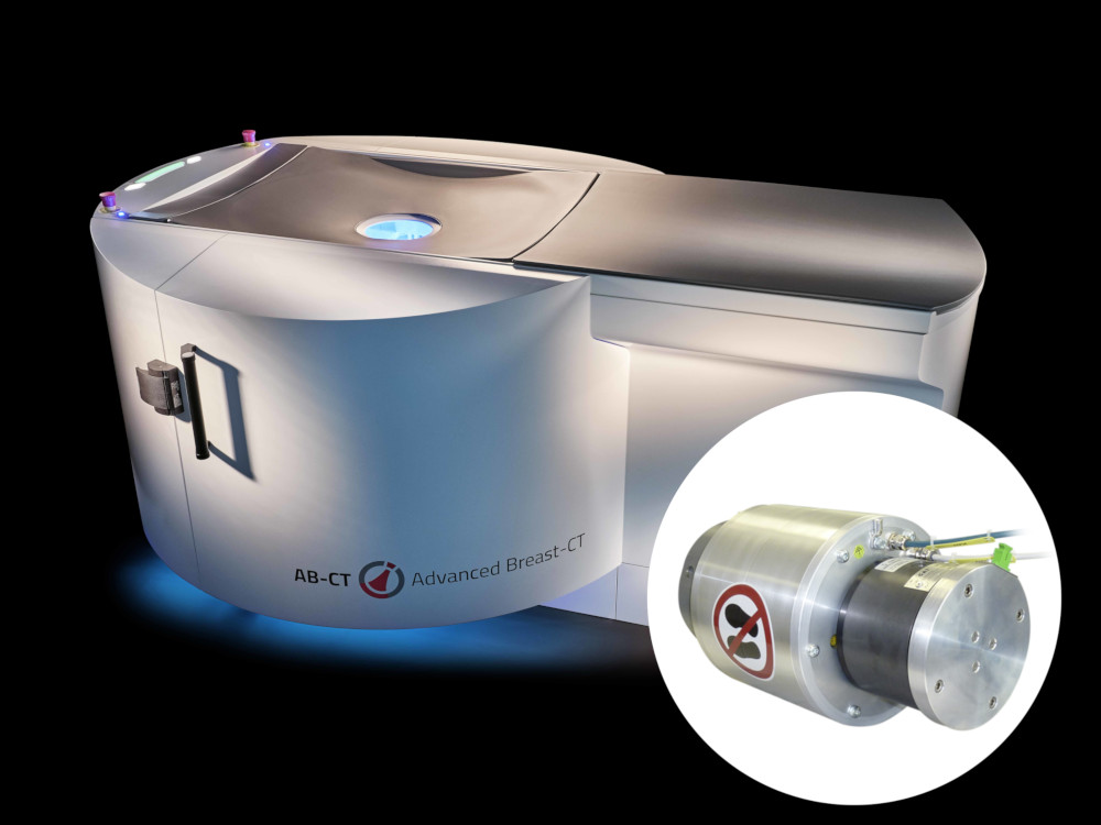
Offering an alternative solution, Moog GAT has partnered with Advanced Breasts GmbH to chart a new path in breast exams. The world’s first breast CT scanner based on spiral CT and photon counting technology has been developed. Moog GAT's customized solution incorporates slip rings, that utilizes gold/gold transmission technology for power supply, rotary unions that transfer coolant to the CT unit, and fiber optic rotary joints (FORJ) that assist in trasmitting the imaging data. The innovative nu:view scanner design places high demands on our installed components, as fast data transmission is required to process the data volume of the high-resolution 3D images.
Without compromizing performance, Moog GAT's experience enabled a compact design to be developed by cleverly arranging the components used from different technologies. This was implemented by positioning the fiber-optic rotary joint within the hollow shaft of the rotary union. This allowed the device to have a low overall height, allowing for easy accessibility for patients using the CT scanner.
A Step Above The Rest
The comfort offered to patients by this new breast scanner should be clearly emphasized, as there is no need for any compression of the breast during the scan. The patient lies prone on the surface of the nu:view and places her breast comfortably in the opening provided. The scan of the breast is performed at a low radiation dose and takes only 7 - 12 seconds. During the examination, up to 12,000 projections are created, allowing nu:view to image all parts of the breast to create a true 3D image without superimposition of multiple images.
Due to its comparatively compact size, the nu:view is not only reserved for radiology clinics, but can also be used in specialist practices. This high-tech imaging system is already being used successfully in several institutions. At the University Hospital Zurich alone, more than 3,000 patients have been examined to date.